Clinical Research and Clinical Case Reports
OPEN ACCESS | Volume 7 - Issue 1 - 2026
ISSN No: 2836-2667 | Journal DOI: 10.61148/2836-2667/CRCCR


S.Bouabdella 1, A.Khouna1, S.Dikhaye1,2, N.Zizi1,2
1Department of Dermatology, Venereology and Allergology, CHU Mohammed VI, Oujda, Morocco
2Laboratory of epidemiology, scientific research and public health.
Faculty of Medicine and Pharmacy, Mohammed Premier University
*Corresponding authors: Sara Bouabdella, Department of Dermatology, Venereology and Allergology, CHU Mohamed VI Oujda, Morocco.
Received: June 17, 2021
Accepted: June 26, 2021
Published: June 29, 2021
Citation: S.Bouabdella, A.Khouna, S.Dikhaye, N.Zizi. “Coral Dermatitis Induced by Moorish Bath: A Case Report”. Clinical Research and Clinical Case Reports, 1(4); DOI: http;//doi.org/04.2021/1.1020
Copyright: © 2021 Sara Bouabdella. This is an open access article distributed under the Creative Commons Attribution License, which permits unrestricted use, distribution, and reproduction in any medium, provided the original work is properly cited.
Corals can elicit both toxic and allergic reactions upon contact with the skin. Clinical presentations vary depending on whether the reaction is acute, delayed, or chronic. Literature concerning cutaneous reactions to corals and other Cnidarians is scarce. Here, we report a case of coral dermatitis after Moorish bathing.
Introduction
The Moorish bath is part of the cultural habits of Moroccans. However, it has several effects on the skin. Corals are known to produce a toxic substance which when comes in to contact with human skin may elicit hypersensitive reactions. We report a rare case of coral dermatitis in an old man with a history of Moorish bathing before being presented to the hospital with skin rashes.
Case Report
A 70-year-old otherwise healthy man presented with a seven-day history of
a slightly pruritic, well-demarcated, erythematous, serpiginous plaque on his thorax developed after going to the Moorish bath.
He reported initially feeling a stinging and burning sensation in his thorax, which was followed by the development of a reddish eruption.
Physical examination revealed a reticulated, well-demarcated, erythematous and serpiginous plaque of his thorax (Figure1). No lymphadenopathy or systemic symptoms were present. The patient had no known allergies.
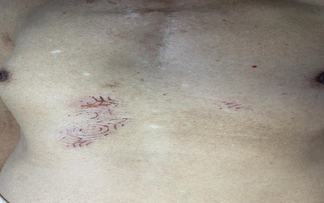
Figure 1: A well-demarcated, erythematous, serpiginous plaque on the thorax
Based on his history and the clinical findings, coral contact dermatitis was diagnosed.
The patient was prescribed a category II topical steroid to apply to the affected area twice daily. The rash and associated pruritus resolved over the following two weeks (Figure 2).
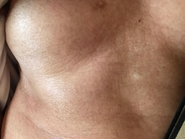
Figure2: The patient after 2 weeks
Discussion
The Moorish baths use water from wells and corals can thrive and live in wells. Along with a variety of aquatic animals such as jellyfish, sea anemones, and hydroids, corals belong to the phylum Cnidaria. Species in this phylum are characterized by the presence of a group of specialized organelles known as cnidocytes. Of these organelles, the nematocyst, which is located in the tentacles of coral, has been implicated in local and systemic reactions in humans as a result of toxic discharge composed of catecholamines and histamines [1].
Coral dermatitis is a skin condition caused by corals [2]. Corals are marine invertebrates that belong to the phylum Cnidaria, which also includes anemones, jellyfish, sea pens and hydra. There are some species of coral that contain nematocysts, a type of organelle [3]. The nematocyst releases its toxins including histamine, histamine liberators, quaternary ammonium compounds, proteins, 5-hydroxytryptamine, and catecholamines [4]. It is known to cause both acute and delayed type hypersensitive/allergic reaction, resulting in skin lesions in humans, very similar to contact dermatitis [2].Coral dermatitis commonly manifests as an acute reaction occurring immediately or within several hours of contact with a coral and typically resolves within several hours to days [4]. The skin lesions present as urticarial or vesicular-bullous plaques, firm, and localized papules, immediately or after hours of exposure to the corals. In some people, superficial epithelioid granulomas were also noted [2]. Delayed hypersensitivity reactions occur less commonly than acute reactions and are triggered by a sequestered antigenic component of the nematocyst [4]. A delayed reaction typically develops over several days to weeks. It is characterized by localized, firm, erythematous papules or plaques and sometimes later by erythematous lichenoid papules or plaques [4].
Nematocyst can also remain harmlessly deposited on the skin for a time, only to erupt later when activated by a specific stimulus. In this case, however, the patient complained of itching for a few minutes immediately after contact with the coral. In addition, the venom may be responsible for extra cutaneous manifestations of immediate allergy such as asthma, but also for delayed hypersensitivity reactions [5]. There are several reports of coral dermatitis in the literature. Most cases have been attributed to the hypersensitive reaction elicited by the immune system against toxic substances produced by the corals [2]. Lesions typically resolve within several weeks of onset. Treatment with oral antihistamines and topical corticosteroids may accelerate time to resolution and reduce symptom burden [1].
Conclusion
Envenomation from corals can be divided into acute and delayed hypersensitivity reactions. Acute reactions are more common and usually occur immediately or within several hours after exposure [3]. Our observation is characterized by the absence of 3 elements normally reported: prior traumatic contact with coral, envenomation erythema, or a fortiori persistent visible coralline material in the wound.


Open Access By Aditum Open Access Journals id licensed under Creative Commons Attribution 4.0 International License. Based On a Work at aditum.org