Clinical Case Reports and Clinical Study
OPEN ACCESS | Volume 13 - Issue 1 - 2026
ISSN No: 2766-8614 | Journal DOI: 10.61148/2766-8614/JCCRCS


Mijo Golemac1, Marija Definis2, Josko Petricevic1*
1Department of Pathology, Citology and Forensic Medicine, University Hospital Center Mostar, Mostar, Bosnia and Herzegovina.
2Department of Pathology, Forensic Medicine and Citology, University Hospital Center Split, Split, Croatia.
*Corresponding Author: Josko Petricevic, Department of Pathology, Citology and Forensic Medicine, University Hospital Center Mostar, Mostar, Bosnia and Herzegovina.
Received: January 22, 2023
Accepted: February 27, 2023
Published: July 04 , 2023
Citation: Mijo Golemac, Marija Definis, Josko Petricevic (2023) “Case Report: Incarcerated Recurrent Obturator Hernia with Richter Component Superficial spreading squamous cell carcinoma in-situ of uterine cervix involving the endometrium: report of a case.”, Clinical Case Reports and Clinical Study, 1(9); DOI: http;//doi.org/01.2023/1.160.
Copyright: © 2023 Josko Petricevic. This is an open access article distributed under the Creative Commons Attribution License, which permits unrestricted use, distribution, and reproduction in any medium, provided the original work is properly Cited.
Superficial spreading squamous cell carcinoma in-situ of uterine cervix involving the endometrium without invading underlaying myometrium, is rare phenomenon. In some very rare cases except endometrium, fallopian tubes and ovaries are also involved. Even the primary squamous carcinoma of endometrium is very rare, in most cases it is result of up stream spred of carcinoma of cervix. Some authors reported that post-radiation therapy of cervical carcinoma contributed in this kind of pattern involving endometrial surface. Our patient is one of rare reported cases in wich in-situ carcinoma of cervix superfitially had spreaded to endometrial surface, without invasion of underlaying myometrium.
Introduction
Cervix is common site of sqamous cell carcinoma, but primary squamous cell carcinoma of the endometrium is very rare condition. Secondary involment by squamous cell carcinoma of cervix is more common and such involment usually occurs via lymphatic rout or by deep myometrial spread (1). Very rare is it's superfitial extension without invasion that was first describe by Cullen (2) in 1900. It can spread further and involving both the fallopian tubes and ovaries wich is extremely rare (3). In last 100 years there were at least 15 simillar reports about this kind of phenomenon wich is connected with post radiation therapy (1,2,3). We report a case of squamous cell carcinoma in-situ of cervix with superfitial extension involving complete endometrium surface, without invasion of underlaying myometrium and no extension to both fallopian tubes and ovaries. This is an rare phenomenon, with less than 10 cases reported so far in the literature.
Case report
A 63-year-old, housewife, presented with postmenopausal spotting for the past 3 months. She was referred to University Clinical Hospital Mostar by a private gynecologist for further treatment. The patient had been menopausal for the past 14 years. A cone biopsy was performed, which on histological examination showed cervical intraepithelial neoplasia III/CIS (carcinoma in-situ). Invasion could not be excluded. After that the patient underwent total abdominal hysterectomy with bilateral salphingo-oophrectomy, and a specimen was submitted for histopathological evaluation.
Pathology findings
Uterus 12x5x4 cm with tubes an ovaries showing small cervix without macroscopically evident tumour mass. Microscopically, both ectocrvical and endocervical epithelium was replaced by malignant squamous cells that were tightly packed and disorganizated showing high nuclear-citoplazmatic ratio and numerous mitotic figures. This change was also seen in endocervical glands, and had superfitially spreaded to the entire endometrium without invading underlaying myometrium (Figure 1). Immunohistochemical marker p16 was used to confirm the origin of malignant squamous cells, showing strong and diffuse cytoplasmic and nuclear expression (Figure 2). The reast endometrial glands were atrophy apeareance with one atrophic polyp of endometrium, mesuring 1,1cm in diametar. In myometrium we found one leiomyoma mesuring 1,5cm in diametar. Fallopian tubes and ovaries shown no pathological findings.
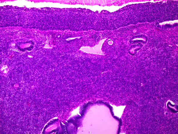
Figure 1. Malignant squamous cells had superfitially spreaded to the entire endometrium without invading underlaying myometrium.
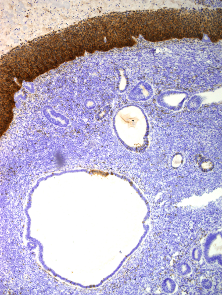
Figure 2. Immunohistochemical marker p16 showing strong and diffuse cytoplasmic and nuclear expression.
Discussion
Altough squamous carcinoma of uterine cervix is common condition, endometrial squamous cell carcinoma is very rare in case (3). Squamous cell carcinom of endometrium can arise from two sources. First one is proposed by Baggish and Woodruff (4) who suggested that primary squamous carcinoma of endometrium probably arise in process of squamous metaplasia. To confirm this diagnosis Fluhmann (5) established three further criteria: no co-existing endometrial carcinoma, no demonstrable connection between the endometrial tumour and the stratified squamous epithelium of cervix, and third one no primary cervical carcinoma. The second mechanism of how squamous carcinoma can be found in endometrium is direct extension from squamous carcinoma of uterine cervix. Biology of this kind of tumour is commonly agrresive showing deep myometrial invasion and dissemination trough lymphatics. Superficial spreading squamous cell carcinoma in-situ of uterine cervix involving the whole surface endometrium can occur very rarely. In our patient this kind of spreading pattern was seen. Microscopically, entire endometrial surface was coverd with malignant squamous epithelium showing no invasion of underlaying myometrium. Both fallopian tubes were lined by normal ciliated columnar epithelium and both ovaries shown no pathological findings. Simmilar to our case there were five cases reported (6,7,8). All of them reported in-situ fashion squamous carcinoma of uterine cervix with superfitial spreading and replacing whole surface endometrium without macroscopic and microscopic signs of invasion of underlaying myometrium. Also in these cases both fallopian tubes and ovaries were without pathological findings. Simillar to theese cases, some authors reported patients hwo have had this pattern also involving falopian tubes (9,10) and even ovaries (3). In some cases authors reported that this pattern was discovered few years after radiation therapy of squamous cell carcinoma of the cervix (9,10). Even there were cases reported considering primary squamous cell carcinoma of endometrium (11) it is obvius that in our case mechanism of superfitial spreding carcinoma of cervix resulted in replacement of columnar epithelium of whole surface endometrium.


Open Access By Aditum Open Access Journals id licensed under Creative Commons Attribution 4.0 International License. Based On a Work at aditum.org