Clinical Case Reports and Clinical Study
OPEN ACCESS | Volume 13 - Issue 1 - 2026
ISSN No: 2766-8614 | Journal DOI: 10.61148/2766-8614/JCCRCS


Andrés Mauricio Camacho Montaño1*, Reinaldo Niño Alba2, María Camila Cetina Grajales3
1 Medical Doctor, Gynecology and Obstetrics Specialist, Universidad del Rosario, Bogotá (Colombia). Maternal-fetal medicine specialist, Sanitas University, Bogotá.
2 Medical Doctor, Gynecology and Obstetrics Specialist, National University, Bogotá (Colombia).
3 Medical Doctor, Universidad Javeriana. Clinica de la Mujer . Bogota (Colombia).
*Corresponding author: Andrés Mauricio Camacho Montaño. Medical Doctor, Gynecology and Obstetrics Specialist, Universidad del Rosario, Bogotá (Colombia). Maternal-fetal medicine specialist, Sanitas University, Bogotá.
Received: January 17, 2021
Accepted: January 27, 2021
Published: February 02, 2021
Citation: Andrés Mauricio Camacho Montaño, Reinaldo Niño Alba, María Camila Cetina Grajales. “Dorsolumbosacral Agenesis. Case Report and Literature Review.”. J Clinical Case Reports and Clinical Study, 1(2); DOI: 10.61148/2766-8614/JCCRCS/018
Copyright: © 2021 Andrés Mauricio Camacho Montaño. This is an open access article distributed under the Creative Commons Attribution License, which permits unrestricted use, distribution, and reproduction in any medium, provided the original work is properly cited.
Objetives: report a case of dorsolumbosacral agenesis and make a systematic review of the literature focused on prenatal diagnosis.
Materials And Methods: we report a case of a 32 year old pregnant woman, with a 30 week pregnanacy, without prenatal care, the fetus is diagnosed with dorsolumbosacral agenesis. The mother request voluntary termination of pregnancy. A systematic review of the literature focused on prenatal diagnosis of thos condiction is performed.
Results: we found 50 papers, 6 met the inclusión critiria. Three of them with prenatal diagnosis. In the first case the diagnosis was made at 13 weeks of gestation and termination of preganancy was requested. In the second case corresponded an biamniotic bicorial twin preganancy. One normal fetus and one with dorsolumbosacral agenesis. The diagnosis was made in the second trimester. The pregnancy continued until 34 week of gestation and the afected neonate had perinatal death. The third case, the diagnosis was made at 18 weeks, a newborn of 2990 gr was born at 37 weeks of gestation.
Conclusion: dorsolumbosacral agenesis is a very severe form of caudal regresión syndrome, with only a few cases reportted in the literature. To the best of our knowladge this is the fourth case reported with prenatal diagnosis.
Introduction
Dorsolumbosacral agenesis is the most severe form of craniocaudal regression syndrome [1]. Caudal regression syndrome has a low incidence, reported 1 in 10,000 births [2] and lumbosacral dysgenesis has only a few cases reported in the literature.
Craniocaudal regression syndrome is a clinical syndrome characterized by malformations of the spine, spinal cord, and lower extremities [2]. Dorsolumbosacral agenesis is characterized by agenesis of the sacrum, lumbar vertebrae and one or more thoracic vertebrae [1] [3].
The etiology of craniocaudal regression is not well clarified, it is known that an alteration in gastrulation occurs before 28 days of gestation [4]. Genes as HLXB9 CYP26A1, F186L and C358R are involved in retinoic acid homeostasis, and have been involved in the pathophysiology of this condition without conclusive data [5] [2]
The main related risk factor is diabetes, which increases the risk 200 times, but drugs such as lithium, sulfonamides or agents such as radiation and extreme temperatures have been also linked with this condition [5]
The diagnosis can be made prenatally by ultrasound, and there are reports of cases reported from the first trimester [6], complementary techniques such as magnetic resonance are very useful to confirm the diagnosis [7] [8].
The prognosis will depend on the extent of the neurological lesion and the associated malformations. It is common to find alterations of the musculoskeletal system, as well as gastrointestinal and urinary [5] [2]. Intelligence is generally normal, but they require long-term neurological, renal, and orthopedic management [4].
Dorsolumbosacral agenesis is a pathology that is little described in the literature, which makes prenatal counseling very difficult. Our objective is to report a case and review the literature focused on the prenatal diagnosis .
Case Report
In August 2019, a 32-year-old woman, migrant from Venezuela, who has lived in Colombia for 1 year, without health care insurance . she went to the emergency unit of Hospital La Victoria; third-level hospital in the city of Bogotá, Colombia.
She had pelvic pain, with a gestation of 28.1 weeks. Medical history: type 2 diabetes mellitus diagnosed 3 years ago, under management with glibenclamide, but she discontinued it, 28 months ago because monetary problems, at the time of the visit without any medical management.
Gynecological history: 4 pregnancies, 2 deliveries, 2 caesarean section, an 1 abortion. Caesarean section for cephalopelvic disproportion 15 years ago and iterative caesarean section 9 years ago. Early abortion 1 year ago. The pregnancy was planned an desired . No prenatal care.
On physical examination: heart rate: 80 bpm, respiratory rate 20rpm, blood pressure 110 / 70mmHg, uterine height: 30cm, fetal heart rate 133bpm, rest of the physical examination within normal limits.
Paraclinical tests: hemogram: hemoglobin 10.4 mg / dl, hematocrit 30. Preprandial 153 and postprandial glycemia 213. Creatinine 0.38 mg / dl. Normal transaminases. Positive treponemal test.HIV negative. Hepatitis B: negative.
Obstetric ultrasound: single fetus breech presentation. In the anatomical evaluation: agenesis of the last dorsal vertebra, all the lumbar vertebrae, of the sacrum andthe coccyx. Figure 1 and 2. . Heart disease: balanced complete atrioventricular canal and bilateral clubfoot. Polyhydramnios: larger pocket 9,3 cm. Posterior placenta with normal characteristics.
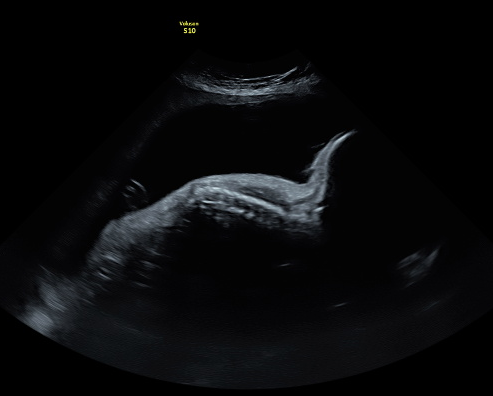
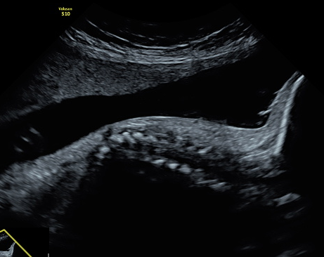
a. Sagittal section of the column b. zoom of the thoracic portion of the spine.
Figure 1. Ultrasound at 30 week.
She was hopsitalized to reach metabolic control. Penicillin and insulin was started, witht a total dose of 0.6 U / kg / day. After three days of hospitalization, metabolic control was achieved.
Patient requests voluntary termination of pregnancy, is assessed by social work, psychology and gynecology group for termination of pregnancy. Informed consent was signed. A fetal asystole induction procedure was performed with the application intracardiac potassium chloride. Procedure withhad no complication., cesarean section was performed. A female fetus of 1500 g, and 28 cm height . X-ray is taken (figure 2). Family did not authorize autopsy.
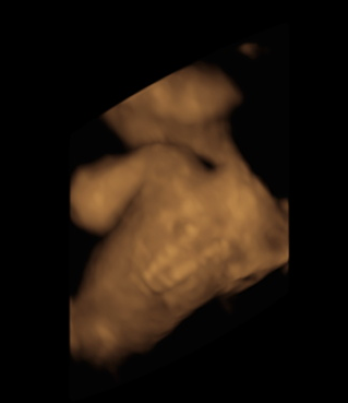
the absence of the lumbar region of the spine is observed
Figure 2. 3D reconstruction
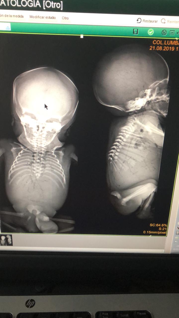
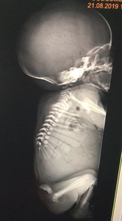
Postmortem radiography. Agenesis of the last thoracic vertebra and lumbosacral vertebrae. a. anteroposterioir view. b. lateral view
Figure 3. Postmortem X- Ray.
The evolution of the puerperium is satisfactory, glucometry in goals and she was sent home, after two days.
Ethical aspects: Authorization was requested from the patient with written informed consent, precautions were taken to guarantee the confidentiality of the information and the anonimity of the patient; the photographic record were taken
by the authors.
Materials And Methods
In order to answer the question of how many cases of dorsolumbosacral agenesis have been diagnosed prenatally, a systematic review of the literature was carried out, with the following mesh terms: "dorsolumbosacral agenesis", "prenatal diagnosis", "caudal regression syndrome". The following databases were searched: Medline via pubmed, and LILIACS. The search was limited to articles published in English or Spanish. The search was limited to the last 30 years. The search was completed in the following databases: OVID, UpToDate and in academic google. A snowball technique was performed by searching the references of the articles. The three investigators carried out the search independently and at the end a reconciliation was made between all authors.
Results
50 related articles were found, of these five fulfilled the inclusion criteria. 5 articles on dorsolumbosacral agenesis (Table 1). One of the articles reported two cases for a total of 6 reported cases. In two cases the diagnosis was made prenatally. In both cases, associated malformations were found (atrioventricular canal and myeloschisis).
|
Author |
country |
Time of diagnosis |
Weeks of gestation |
observations |
|
Mihmanli 2001 (9) |
Turkey |
Newborn of 3 months. |
Does not apply. |
No follow-up. lost after diagnosis. |
|
Nagy 2008 (3) |
Hungary |
Prenatal |
21weeks. |
Voluntary termination of pregnancy |
|
Luque 2001 (10) |
Chile |
Prenatal |
18 weeks. |
2900g newborn. 13-month follow-up. |
|
Bosemani 2013 (1) |
USA |
Prenatal |
13 weeks |
Early perinatal death. |
|
Szumera 2017 (11) |
Poland |
6 years of life |
Does not apply. |
Stabilization surgery was performed |
|
8 years of life |
Does not apply. |
Stabilization surgery was performed |
Table 1. Cases of dorsolumbosacral agenesis.
In the first case, Mihmanli et al reported a 3-month-old newborn, the last vertebra visualized in the MRI was T9. Patient from a rural area of Turkey, who is diagnosed and an orthopedic management is proposed , but the family did not accept; follow up was not possible [9].
The second case, Nagy et al reported a 37-year-old pregnant woman with type 2 diabetes mellitus. Prenatal diagnosis was made by ultrasound at 21 weeks of gestation. The last vertebra visualized was T5, in addition, ventricular communication and a single umbilical artery were found. The patient requested voluntary termination of pregnancy [3].
The third case, Loque et al reported, a 34-year-old pregnant woman, with type 2 diabetes mellitus, diagnosis made by ultrasound at week 18. Agenesis of the last 4 dorsal vertebrae and the lumbosacral spine was documented. A 37-week-old newborn weighing 2990 grams was obtained with 13-month follow-up in rehabilitation management [10].
The fourth case, Bosemani et al reported a 40-year-old pregnant woman, with a history of thyroid cancer treated surgically, without a history of diabetes. At week 13, a diagnosis of biamniotic bicorial multiple gestation with lumbosacral agenesis of fetus A and dorsal myelomennigocele was made. fetus B normal. In week 21, an MRI was performed in week 21 evidence of agenesis of the last thoracic vertebrae, lumbar vertebrae and sacrum. In addition to thoracic myelomengocele, Chiari II malformation and bilateral clubfoot. Termination of the pregnancy of fetus A was proposed. The patient decides to continue the pregnancy, at week 33 she presented rupture of membranes. The neonate with the malformations died 6 days after birth, the healthy neonate has a satisfactory evolution [1].
Szumera et al [11], reported two pediatric cases. The first case a 6-year-old girl with agenesis of the last dorsal vertebra, the lumbar vertebrae and the sacrum; and the second case an 8-year-old boy with agenesis of the last three dorsal vertebrae,lumbar vertebrae and sacrum. The two cases underwent a successful spinal fixation orthopedic procedure for stabilization.
Conclusions
Dorsolumbosacral agenesis is an extreme rare presentation of the craniocaudal regression syndrome, with a very low incidence. The diagnosis can be made prenatally by ultrasound, complementary tests such as resonance can be useful.
It is important for the clinical practice to understand this pathology for better prenatal counseling to parents and more informed decision-making.
Authors 'contribution:
All authors participated equally in the preparation of the document, from its conception and design to the acquisition of the information, review of the intellectual content and approval of the version sent to the editorial process.
Financing: own resources.
Conflicts Of Interest: None.


Open Access By Aditum Open Access Journals id licensed under Creative Commons Attribution 4.0 International License. Based On a Work at aditum.org