Clinical Case Reports and Clinical Study
OPEN ACCESS | Volume 13 - Issue 1 - 2026
ISSN No: 2766-8614 | Journal DOI: 10.61148/2766-8614/JCCRCS


Nanda Rachmad Putra Gofur 1*, Aisyah Rachmadani Putri Gofur 2, Soesilaningtyas 3, Rizki Nur Rachman Putra Gofur 4, Mega Kahdina 4, Hernalia Martadila Putri 4,
1Department of Health, Faculty of Vocational Studies, Universitas Airlangga, Surabaya, Indonesia
2Faculty of Dental Medicine, Universitas Airlangga, Surabaya, Indonesia
3Department of Dental Nursing, Poltekkes Kemenkes, Surabaya, Indonesia
4Faculty Of Medicine, Universitas Airlangga, Surabaya, Indonesia
*Corresponding author: Nanda Rachmad Putra Gofur, Department of Health, Faculty of Vocational Studies, Universitas Airlangga, Surabaya, Indonesia
Received: March 01, 2021
Accepted: March 25, 2021
Published: April 05, 2021
Citation: Putra Gofur N R, Putri Gofur A R, Soesilaningtyas, Rachman Putra Gofur R N , Kahdina M, “ Neurotheology: Insights on the Relationship between the Brain and Religion Through the Life and Ministry of St. Paul the Apostle”. Clinical Case Reports and Clinical Study, 3(2); DOI: 10.61148/2766-8614/JCCRCS/044
Copyright: © 2021 Nanda Rachmad Putra Gofur. This is an open access article distributed under the Creative Commons Attribution License, which permits unrestricted use, distribution, and reproduction in any medium, provided the original work is properly cited.
Melanin granules are basically inside the keratinocytes, melanin granules will collect in the supranuclear area of the cytoplasm so that the nucleus will be protected from the effects of damaging solar radiation. Although melanocytes form melanin, but those that function as a depot and contain more melanin pigment than melanocytes are epithelial cells. In keratinocytes, the melanin granules are fused with lysosomes. This is why melanin disappears in the upper cells. In the interaction process between keratinocytes and melanocytes, which results in skin pigmentation, the most important factors are the speed of melanin granule formation in melanocytes, transfer of granules into keratinocytes, and the final disposition of granules by keratinocytes. Human skin color is determined by various pigments. Those that play a role in determining skin color are carotene, melanin, oxyhemoglobin and reduced forms of hemoglobin, the most important of which is the pigment melanin. Melanosis is a disorder in the process of forming the skin's melanin pigment. First is hypermelanosis (melanoderma) when the production of the pigment melanin increases, can be caused by increased melanocyte cells or only increased melanin pigment. Conversely, leukoderma can be caused by a reduction in the amount of melanin pigment or a decrease or absence of melanocyte cells. Hypermelanosis of both types can result from three factors: genetic, hormonal (as in Addison's disease), when it is caused by increased circulating pituitary melanotropic hormone, and UVR. Aims of this article is to review melasma, clinical manifestation and management.
Discussion : Melasma is acquired hypermelanosis that is generally symmetrical in the form of a light brown to dark brown patch of macules, affecting areas exposed to ultraviolet light with predilection sites on the cheeks, forehead, upper lip, nose and chin. Melasma can affect all races, especially people living in tropical areas. Melasma is mainly found in women, although it is also found in men (10%). In Indonesia, the ratio of cases of women and men is 24: 1. This is especially evident in women of childbearing age with a history of direct sun exposure. Most incidence is at the age of 30-44 years.Melasma treatment requires a long time, regular control and good cooperation between the patient and the doctor who handles the case. Most of these patients seek cosmetic reasons. Treatment and skin care must be carried out regularly and because melasma is chronic residual disease. Principle of treatment is a causal treatment, so it is important to find the etiology. Prevention against the onset or gaining weight and recurrence of melasma is protection against sunlight. Patients are required to avoid direct exposure to ultraviolet rays, especially between 09.00 and 15.00. preferably if you leave the house using a wide umbrella or hat.
Conclusion : Risk factor of melasma is women with a history of direct sun exposure. Melasma lesions are defined light brown or dark brown macules with irregular edges, often on the cheeks, and nose, known as a malar pattern. Melasma treatment requires a long time, regular control and good cooperation between the patient and the doctor who handles the case. Principle of treatment is a causal treatment, so it is important to find the etiology.
Introduction
Melanin granules are basically injected into keratinocytes. Once inside the keratinocytes, melanin granules will collect in the supranuclear area of the cytoplasm so that the nucleus will be protected from the effects of damaging solar radiation. Although melanocytes form melanin, but those that function as a depot and contain more melanin pigment than melanocytes are epithelial cells. In keratinocytes, the melanin granules are fused with lysosomes. This is why melanin disappears in the upper cells. In the interaction process between keratinocytes and melanocytes, which results in skin pigmentation, the most important factors are the speed of melanin granule formation in melanocytes, transfer of granules into keratinocytes, and the final disposition of granules by keratinocytes. A feedback mechanism is likely between melanocytes and keratinocytes1.
Melanocytes can be easily seen by incubating epidermal fragments in dopa. These compounds are converted into dark brown melanin deposits in melanocytes, a reaction catalyzed by the enzyme tyrosinase. In this way, the number of melanocytes in the epidermal area can be counted. The study demonstrated that these cells were not randomly distributed among keratinocytes; however, there is a dispersal pattern called the melanin-epidermal unit. In humans, the ratio of dopa-positive melanocytes to keratinocytes in the stratum basale is constant in each region of the body, but varies from region to region. For example, there are about 1000 melanocytes / mm2 in the skin of the thigh, and 2000 / mm2 in the skin of the scrotum. The number of melanocytes per unit area was not influenced by sex or race, and differences in skin color were mainly due to differences in the number of melanin granules in keratinocytes2.
Human skin color is determined by various pigments. Those that play a role in determining skin color are carotene, melanin, oxyhemoglobin and reduced forms of hemoglobin, the most important of which is the pigment melanin. Melanosis is a disorder in the process of forming the skin's melanin pigment. First is hypermelanosis (melanoderma) when the production of the pigment melanin increases, can be caused by increased melanocyte cells or only increased melanin pigment. Conversely, leukoderma can be caused by a reduction in the amount of melanin pigment or a decrease or absence of melanocyte cells. Hypermelanosis of both types can result from three factors: genetic, hormonal (as in Addison's disease), when it is caused by increased circulating pituitary melanotropic hormone, and UVR. Aims of this article is to review melasma, clinical manifestation and management3.
Discussion
Melasma is acquired hypermelanosis that is generally symmetrical in the form of a light brown to dark brown patch of macules, affecting areas exposed to ultraviolet light with predilection sites on the cheeks, forehead, upper lip, nose and chin. Melasma can affect all races, especially people living in tropical areas. Melasma is mainly found in women, although it is also found in men (10%). In Indonesia, the ratio of cases of women and men is 24: 1. This is especially evident in women of childbearing age with a history of direct sun exposure. Most incidence is at the age of 30-44 years4.
Abnormalities can affect pregnant women, women using contraceptive pills, cosmetic users, drug users, etc. Until now it is still needed further studies. The causative factors that are thought to play a role in the pathogenesis of melasma are5:
1. Ultraviolet light. This spectrum of sunlight destroys the sulfhydryl groups of the epidermis which are Tyrosinase enzyme inhibitor by binding to Cu ions from the enzyme. Ultra violet light causes the tyrosinase enzyme to no longer be inhibited, thus spurring the melanogenesis process.
2. Hormones. For example, estrogen, progesterone and MSH play a role in the development of melasma. In pregnancy, melasma usually extends in the third trimester. In contraceptive pill users, melasma appears within 1 month to 2 years after starting the pill.
3. Medicine. For example diphenyl hidantoin, mesantoin, chlorpromacin, cytostatic, and minocycline can cause melasma. This drug is piled in the upper dermis layer and cumulatively can stimulate melanogenesis.
4. Genetic. 20-70% of family cases are reported
5. Race. Melasma is often found in the Hispanic and dark skin groups
6. Cosmetics. The use of cosmetics that contain perfume, dyes, or certain ingredients can cause photo sensitivity which can cause hyperpigmentation on the face if exposed to sunlight.
7. Idiopathic
Classification of melasma
There are several types of melasma in terms of clinical features, histopathological examination, and examination with wood rays. Based on the clinical picture6:
1. The centro-facial form includes the forehead, nose, cheeks, medial part, under the nose and chin
2. The malar shape includes the lateral nose and cheeks.
3. Mandibular shape covers the mandibular area
Based on inspection with wood rays
1. Epidermal type, melasma is clearer on wood light than ordinary light
2. Dermal type, with wood light there is no contrasting color compared to ordinary light
3. Mixed type, some locations appear more clearly while others are not clear
4. The type is difficult to assess because of the dark color of the skin, with wood light the lesion becomes unclear, while with ordinary light it is clearly visible. The difference between these types is very significant in the treatment administration, the dermal type is more difficult to treat than the epidermal type.
Based on histopathological examination7:
1. Melasma epidermal type, generally brown in color, melanin is mainly found in the basal and suprabasal layers, sometimes throughout the stratum corneum and stratum spinosum.
2. Dermal type Melasma, bluish brown in color, there are macrophages burrowing around the blood vessels in the upper and lower dermis, in the upper dermis there are foci of infiltrates
The pathogenesis of melasma is much remains unknown. Many factors are involved in this process, including:
a. Increased production of melanosomes due to hormones and due to ultraviolet light. This increase in melanosomes can also be caused by pharmacological substances such as silver and psoralen.
b. Inhibition in malphigian cell turnover, this situation can occur due to cytotoxic drugs.
Clinical Manifestation of Melasma
Melasma lesions are defined light brown or dark brown macules with irregular edges, often on the cheeks, and nose, known as a malar pattern. The mandibular pattern is found on the chin, while the centrofacial pattern is found on the temples, forehead, eyebrows and upper lip. Grayish or bluish color, especially in the dermal type8.
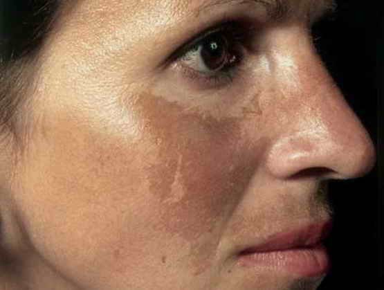
Figure 1. Hyperpigmented macules seen on the cheeks, nose, and upper lip.
Through histopathological examination, there are 2 types of hypermelanosis, first epidermal type, melaninn mainly present in the basal and suprabasal layers, sometimes throughout the stratum spinosa to the stratum corneum; cells that contain dense melanin are melanocytes, basal and suprabasal layer cells are also present in keratinocytes and cells of the startum corneum. Then dermal type, there are macrophages with melanin around the blood vessels in the upper and lower dermis; in the upper and lower dermis; in the upper dermis there are foci of infiltrates9.
Electron microscope examination, The ultrastructure of melanocytes in the basal layer suggests increased melanost activity. Examination with wood rays divided into 4 type as epidermal type: the color of the lesion appears more contrasting, dermal type: the color of the lesion does not increase in contrast, mixed type there are lesions that increase in contrast or not and indistinct type: with wodd light the lesion becomes indistinct, whereas with ordinary light it is clearly visible. The diagnosis of melasma is only made by clinical examination. To determine the type of melasma, a wood ray examination is performed, while histopathological examination is only carried out in certain cases10.
Management of Melasma
Melasma treatment requires a long time, regular control and good cooperation between the patient and the doctor who handles the case. Most of these patients seek cosmetic reasons. Treatment and skin care must be carried out regularly and because melasma is chronic residual disease. Principle of treatment is a causal treatment, so it is important to find the etiology. Prevention against the onset or gaining weight and recurrence of melasma is protection against sunlight. Patients are required to avoid direct exposure to ultraviolet rays, especially between 09.00 and 15.00. preferably if you leave the house using a wide umbrella or hat11,12.
Protect the skin by wearing the right sunscreen, both regarding the ingredients and how to use it. Without the use of sunscreen every day treatment is difficult to succeed. It is recommended that you use sunscreen 30 minutes before sun exposure. There are 2 types of sunscreens that are known, namely physical sunscreens and chemical sunscreens. Physical sunscreens are materials that can reflect or scatter ultraviolet; for example titanium dioxide, zinc oxide, kaolin; while the chemical barrier is material that absorbs ultraviolet13,14.
There are 2 types of chemical sunscreens, first containing PABA (paraamino benzoic acid) or its derivatives, for example octil PABA and that does not contain PABA, for example, bensophenone, cinnamic, salicylate, and anthranylate. Eliminating factors that cause melasma, for example stopping the use of contraceptive pills, stopping the use of cosmetics that are colored or containing perfume, preventing drugs such as hydantoin, cytostatics, anti-malarial drugs, and minocycline15,16.
Management of topical treatment could use hydroquinone. Hydroquinone is used at a concentration of 2-5%. the cream is used at night accompanied by the use of a sunscreen during the day. Generally there is improvement in 6-8 weeks and continued for up to 6 months. A side effect is irritant or allergic contact dermatitis. After discontinuation of the use of hydroquinone relapses often occur. Moreover, retinoic acid 0.1% is mainly used as adjunct therapy or combination therapy. The cream is also used at night, because during the day there can be photodegradation. Now retinoic acid is used as monotherapy, and there is significant clinical improvement, although it is slow. Side effects include erythema, desquamation and photosensitation. Last, azelic acid is a safe drug to use. Treatment with azelic acid 20% for 6 months gave good results. The side effects are burning and itching17,18.
Management of topical treatment could use systemic treatment absorbic acid/ vitamin C. Vitamin C has the effect of converting oxidized melanin into reduced melanin, which is brighter in color and prevents melanin formation by converting DOPA kinones into DOPA. Second, alternative is gluthaThe reduced form of glutathione is a sulfhydryl (SH) compound that has the potential to inhibit melanin formation by combining with cuprum from tyrosinase. Another option is chemical peel and laser surgery. Chemical peels can help treat hyperpigmentation disorders. Chemical peeling is done by applying 50-70% glycolic acid for 4 to 6 minutes, done every 3 weeks for 6 times. Before the chemical peel is given 10% glycolic acid cream for 14 days. Laser surgery using a Q-switched ruby laser and an argon laser, relapse may also occur19,20.
Conclusion
Risk factor of melasma is women with a history of direct sun exposure. Melasma lesions are defined light brown or dark brown macules with irregular edges, often on the cheeks, and nose, known as a malar pattern. Melasma treatment requires a long time, regular control and good cooperation between the patient and the doctor who handles the case. Principle of treatment is a causal treatment, so it is important to find the etiology.


Open Access By Aditum Open Access Journals id licensed under Creative Commons Attribution 4.0 International License. Based On a Work at aditum.org