Mohammed Basith 1, Dilip Kumar G2, Krishna Prasad T3, Ameen Hanan 4
1Shri Sathya Sai Medical College and RI, Ammapettai, Kanchipuram Dt-603108.
2Shri Sathya Sai Medical College And Ri, Ammapettai, Kanchipuram DT-603108.
3Shri Sathya Sai Medical College and RI, Ammapettai, Kanchipuram Dt-603108.
4Resident, department of dermatology.
*Corresponding author: Krishna Prasad T, Shri Sathya Sai Medical College and RI, Ammapettai, Kanchipuram Dt- 603108.
Received: June 10, 2024
Accepted: June 15, 2024
Published: June 17, 2024
Citation: Basith M, Dilip Kumar G, Krishna Prasad T, Hanan A. (2024) “Anesthetic Management of a Young Female with Kyphoscoliosis and Severe Pulmonary Hypertension Posted for Appendicectomy- A Case Report.” Anesthesia and Pain Management, 1(1); DOI: 10.61148/2836-077X/APM/002
Copyright: © 2024 Krishna Prasad T. This is an open access article distributed under the Creative Commons Attribution License, which permits unrestricted use, distribution, and reproduction in any medium, provided the original work is properly cited.
Kyphoscoliosis may cause compromise in pulmonary function due to restrictive lung disease, pulmonary hypertension, cardiac compression, and heart failure. There is a decrease in total lung capacity and chest wall compliance. In this case, an 18-year-old female with Kyphoscoliosis was diagnosed with severe pulmonary hypertension and on treatment with diuretics. ECG showed P- Pulmonale, right axis deviation, and right bundle branch block and ECHO revealed severe tricuspid regurgitation with severe pulmonary hypertension, this patient presented with right iliac fossa tenderness and CT revealed inflamed appendix filled with fluid and appendicolith and she was planned for open appendicectomy. our choice of anesthesia was spinal with continuous epidural block and procedure was uneventful. Hence regional anesthesia with careful approach and monitoring can be a useful technique of providing safe and effective anesthesia in patients with kyphoscoliosis with pulmonary hypertension.
kyphoscoliosis; epidural block; pulmonary hypertension
Introduction:
Kyphoscoliosis is defined as lateral and posterior curving of the spine. It can affect the lumbar, thoracic region, or both. It can occur as an isolated idiopathic condition or secondary to numerous disorders such as Marfan syndrome, muscular dystrophy, collagen vascular disorders, and neurofibromatosis [1]. This indicates the importance of pre-operative evaluation to rule out co-existing disorders.
Thoracic spine deformity may cause compromise in pulmonary function due to restrictive lung disease, pulmonary hypertension, cardiac compression, and heart failure. There is a decrease in total lung capacity and chest wall compliance [2]. Thus assessing the degree of curvature, cardiopulmonary signs, and symptoms are essential in these patients.
Possible mechanisms for pulmonary hypertension are hypoxemia and hypoventilation leading to pulmonary vasoconstriction. This leads to right ventricular hypertrophy and failure [3]. Restrictive lung disease, airway management, and cardiopulmonary compromise make general anesthesia complicated, whereas epidural and spinal anesthesia is associated with technical difficulties due to abnormal curvature of the spine.
Here we report a case of a young female with kyphoscoliosis and severe pulmonary hypertension planned for appendicectomy under regional anesthesia.
Case Report:
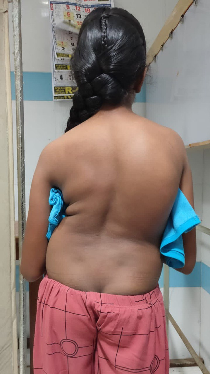
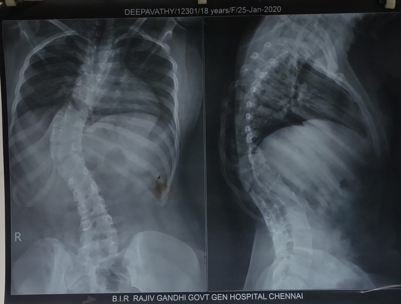
An 18-year-old female with kyphoscoliosis (figure 1), was diagnosed with severe pulmonary hypertension 4 years back. She has been on treatment with T. Furosemide 20mg BD, T. Aldactone 25mg OD, and T. Tadalafil 20mg OD since then. She was admitted to the surgical ward as a case of acute abdomen for 2 days medicated with fluids and analgesics. On clinical examination, she had right upper quadrant and right iliac fossa tenderness. There was no rigidity, guarding, or rebound tenderness. She was febrile with a temperature of 37.6℃. Her blood tests revealed leucocytosis with neutrophilia (82%), anemia with Hb 10mg/dl and CRP 54mg/L. All other preoperative blood investigations were within normal limits. ECG showed P pulmonale, right axis deviation, and right bundle branch block. Chest X-ray showed kyphoscoliosis with bilateral hyperinflated lungs and crowding of the ribs (figure 2). Metabolic equivalence was 25 seconds.
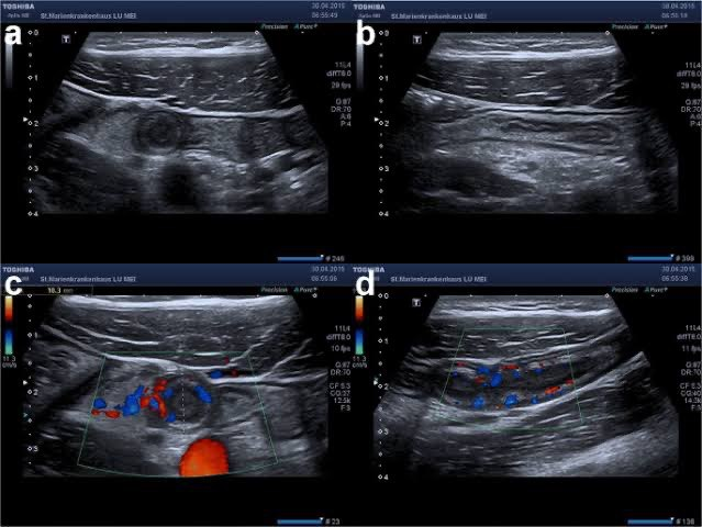
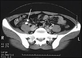
Ultrasound scan of the abdomen revealed a dilated tubular structure with fluid in the right lower quadrant with a blind-end tip, increased echogenicity, and hyperemia of the peri-appendiceal fat with appendicolith (figure 3). CT showed an inflamed appendix filled with fluid and an appendicolith (figure 4). 2D ECHO revealed an ejection fraction of 65%, normal left ventricular systolic function, no regional wall motion abnormalities, dilated right atrium and right ventricle, severe tricuspid regurgitation, and severe pulmonary hypertension. There were no signs and symptoms of congestive cardiac failure. Her pulmonary function tests revealed restrictive pattern.

Figure 1: An 18-year-old female with kyphoscoliosis

Figure 2: Kyphoscoliosis with bilateral hyper inflated lungs and crowding of the ribs

Figure 3: The peri-appendiceal fat with appendicolith

Figure 4: Inflamed appendix filled with fluid and an appendicolith
The patient gave consent for high-risk anesthesia under American Society of Anesthesiologists grade III E. Preoperative parameters were pulse rate 104/min, blood pressure 100/60mmHg, and SPO2 98% in room air. Mallampatti claasification grade was 2. Baseline monitoring of pulse rate, blood pressure, SPO2, capnography, and urine output was done. Our choice of anesthesia in this patient was spinal with continuous epidural block. Under aseptic precautions, the patient in a sitting position, using 18 gauge Tuohy’s needle epidural space was identified using loss of resistance technique in L1-L2 space. The catheter was fixed 7cm at skin level. Spinal anesthesia was given at L3-L4 level with 2.7ml of 0.5% inj. bupivacaine with 0.5ml of inj. fentanyl. Epidural top-up was done using 0.25% bupivacaine after every 45 mins. Analgesia was achieved till T6 dermatome.
Surgically, muscle splitting Lanz incision was made in the right iliac fossa, The appendix was swollen, inflamed, and paracaecal in position. After dividing the mesoappendix, appendicectomy was performed. Surgery proceeded for 3 hours. No significant hemodynamic changes were observed during the surgery. Postoperatively, the epidural was retained for analgesia and was removed on 2nd day. The patient was comfortable throughout the postoperative period.
Discussion:
In kyphoscoliosis, the structural and dynamic counterparts of the spine are disrupted resulting in a restrictive type of lung pathology [4]. The anesthetic and intraoperative management is highly challenging in these patients since they are hemodynamically unstable [5].
The primary goal of anesthetic management in patients with kyphoscoliosis and pulmonary hypertension would be the maintenance of normovolemia, avoidance of hypoxemia, hypercapnia, pain, acidosis, and drug-induced myocardial depression [6]. All these factors contribute to an increase in pulmonary vascular resistance with rapid onset of right ventricular decompensation and sudden cardiac arrest. Thus proper maintenance of intravascular volume, systemic vascular resistance, and preload is necessary [7,8].
Regional anesthesia is a better alternative to general anesthesia in these types of patients as it blocks the pain-induced rise in pulmonary vascular resistance (PVR), avoids the deleterious effect of PEEP on PVR and right ventricular function as well as facing the difficulty of extubation or weaning from ventilation [9].
In contrast, general anesthesia can be preferred in selected patients posted for prolonged or major surgeries and in severe uncontrolled pulmonary hypertension as selective pulmonary vasodilators can be administered through the ventilatory circuits [10].
The apex of the scoliotic curve is where the greatest rotation occurs. In untreated patients, there is a strong linear association between the Cobb angle and vertebral rotation in both the thoracic and lumbar curves. Thus giving a tough job for the anesthetist in performing regional anesthesia. Neural damage, spinal hematomas, post-dural puncture headaches, and infections can arise from conducting neuraxial anesthesia in such a patient with pulmonary vascular changes.
The anesthetist should proceed cautiously in cases with neuromuscular scoliosis if the anatomy is simple and well-understood. However, since these scoliotic curves are sometimes complex, other options for placement or pain management should be carefully considered.
Conclusion:
The decision of choosing which type of anesthesia for a patient becomes technically difficult when both the airway and spine are involved in the disease process. Regional anesthesia with careful approach and monitoring can be a useful technique for providing safe and effective anesthesia in patients with kyphoscoliosis with pulmonary hypertension.