Aditum Journal of Clinical and Biomedical Research
OPEN ACCESS | Volume 8 - Issue 1 - 2026
ISSN No: 2993-9968 | Journal DOI: 10.61148/2993-9968/AJCBR


Michel Leclerc
556 rue Isabelle Romée, 45640 Sandillon France.
Corresponding Author: Michel Leclerc, 556 rue Isabelle Romée, 45640 Sandillon France.
Received: November 10, 2021
Accepted: December 20, 2021
Published: January 03, 2022
Citation: Michel Leclerc. (2022) “ Asterias rubens (Echinoderm) : Evidence of Lymphocytes, Lymphokines and Invertebrate Primitive antibody(IPA).Expected rôle of Tiedemann’s bodies.”, Aditum Journal of Clinical and Biomedical Research, 4(1); DOI: http;//doi.org/01.2022/1.1065.
Copyright: © 2022. Michel Leclerc. This is an open access article distributed under the Creative Commons Attribution License, which permits unrestricted use, distribution, and reproduction in any medium, provided the original work is properly Cited.
The axial organ is considered as ancestral lymphoïd organ. It contains T and B sea star lymphocytes and Phagocytes. It plays a fundamental rôle in the sea star cell-mediated immune responses and humoral immune ones.Asterids belong to Echinoderma(Invertebrates).An expected rôle of Tiedemann’s bodies is evoked.
Introduction :
The position of the echinoderms, in particular, has been a matter of doubt and controversy in the studies of the vertebrate ancestors (Kampmeier : [1])
Sea-stars belong to the class Asteroidea of the phylum Echinoderma : Asterias rubens was studied.
They are marine animals, also named starfishes. They possess a primitive lymphoïd organ :
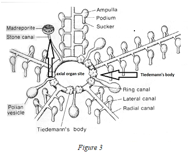
The axial organ which lies along the stone canal, the Tiedemann’s bodies (2 per interradius:total : 8) situated in the ring canal of the oral part .
The structure of the axial organ is glandular, spongy and crossed by connective tissue.
Many follicles of heterogeneous size with clear zones are lined by cellular cords. We find a similar structure inside the Tiedemann’s bodies : they evoke lymph nodes.A schema represents these last ones in Fig.3
The various types of cells present in the axial organ have been studied in our laboratory, by optical and electron microscopy (S.E.M and T.E.M).
Essentially two types of cells are found in T.E.M [2]:
Cells which morphologically resemble mammal lymphocytes ( Fig.1) (Fig .2).

Evidence of Lymphokines :
The method of Leiper and Solomon [3] was used, with some modifications.Cells were separated by nylon wool and were cultured for 24 h at 10° C in the presence of PWM-coated Sepharose beads. Supernatants from these cultures were centrifuged to eliminate beads and debris, sterilized by membrane filtration (0.22-µm Millipore) and concentrated 5- or 10-fold with an ultrafiltration device (Amicon) fitted with « PM-10 Diaflo » membranes. Supernatant controls were obtained from cells cultured with non-coated beads, from cells cultured alone and from beads cultured alone. The supernatants were added to cultures of total axial organ cells which had been started 24 h before, and 24 h later, the cells were harvested and mitogenic stimulation was evaluated by measuring tritiated-H-methyl- thymidine incorporation.
It was found that the the supernatant from the non-adherent cells cultured in the presence of PWM beads stimulated the total axial organ cells. No stimulation was observed in the various controls. This result indicates that non-adherent cells(i-e sea star T lymphocytes) produce a lymphokine mediator which is able to stimulate the total axial organ cells. The active substance was inactivated by trypsin and chemotrypsin, or by heating for 1 h at 70°C, but it resisted at 56°C. [4]
In 1997, cytokines and cytokine-receptors were discovered indirectly by the use of Cytofluorometry [5] and genomic studies [6]
Evidence of Humoral Responses :
Notion of IPA (Invertebrate primitive antibody) :
In a first study(Leclerc, 1973, [7]), it was shown that the Asterina gibbosa axial organ cells, previously injected with horse-radish peroxydase(HRP) were able to react specifically with the same enzyme. This observation was made by T.E.M, using an immunocytochemical technique.A similar result was later observed with the Asterid : Asterias rubens. Since a variety of antigens, were injected in the coelomic cavity, near the axial organ of Asterids, such as : E.coli alkaline phosphatase, bovine serum albumin, rat IgG, human lambda Bence-Jones protein, myoglobin, and more recently, the haptens : TNP and FITC coupled to polyacrylamide beads . Results were summarized in 2012 [8] We identified an invertebrate primitive antibody, characteristics of which were : specificity and structure(2 Ig sites) after its(sea star) immunization to HRP and genomic research [9]: The gene showed a specific immune response to the enzyme HRP after its insertion in an Escherichia coli plasmid [10].Moreover Fab gene , Fc receptor gene,Cr gene and MHC genes were recently discovered [11]
Conclusions :
The general idea that emerges from the experiments reported in the present review is that Echinoderma, as exemplified by the sea star: Asterias rubens, possess an ancestral lymphoïd organ: the axial organ. It appears that all the elements necessary to immune responses ( phagocytic cells, T sea star lymphocytes, B lymphocytes,) are present in this organ which initiates : lymphokines, humoral immune responses (invertebrate primitive antibody : IPA) of the sea star.We suppose now that immune responses occur also from Tiedemann’s bodies which may be compared to Vertebrate lymph nodes.At the begining these structures were considered as enigmatic [12]. Further studies are necessary to confirm or to infirm the idea they are resembling to vertebrate lymph nodes.


Open Access By Aditum Open Access Journals id licensed under Creative Commons Attribution 4.0 International License. Based On a Work at aditum.org